

Please bookmark this page and visit weekly for the latest updates! Rhabdomyosarcoma is a rare cancer of children, adolescents and young adults (and sometimes older adults). There are two major subtypes, alveolar (ARMS) and embryonal (ERMS). Rhabdomyosarcoma is particularly difficult to treat when it has spread to other parts of the body. This process of spreading is called metastasis. Why does rhabdomyosarcoma spread? The answer may involvea protein called, Osteopontin. Osteopontin is made by rhabdomyosarcoma tumor cells and shared in the tissue around the tumor cells. It is thought that bringing blood vessels to the tumor cells is one way in which Osteopontin helps rhabdomyosarcoma to progress … but there may be other aspects to how osteopontin remodels the tissue “micro-environment”, and we aim to understand these changes. This project is currently led by cc-TDI postdoctoral fellow, Dr. Alexandria (Ali) Harrold.
Please check back weekly to see our progress!
update 6/06/2018:
By popular demand, we are switching to less-frequent, more content-rich monthly posts. These will appear as new blog posts on our website. For exclusive! week to week updates of late-breaking results, email molly@cc-tdi.org to be subscribed. Thank you for supporting this rhabdomyosarcoma research!
———————————–
01/03/2018: Defining the role of osteopontin in rhabdomyosarcoma tumor cells involves a bit of ‘push and pull’… that is, taking osteopontin away, then adding it back. Our first approach for ARMS and ERMS has been to take osteopontin away completely, using a technology called CRISPR. To study the effect another way, we are using a technology called shRNA (to turn osteopontin off in a matter of degrees rather than a simple on/off), and we will also turn osteopontin on using cDNA over-expression. These steps take a while… so bear with us as we move this aspect of the project forward! The result of these genetic proof-of-concept studies will be the foundation for developing and testing drugs that aim to have the same effect as the genetic knockdowns.

01/24/2018: This week the Osteopontin project has been a bit like a swimming duck: everything looks calm on the surface but underneath we’re paddling furiously and moving forward. On Monday, Dr. Aykut Uren from Georgetown University visited the lab and we had an very enlivening conversation about Osteopontin. More antibodies arrived and will be tested later this week; we were even able to get some synthetic Osteopontin protein and cells that don’t have Osteopontin to thoroughly evaluate all the antibodies we use. We’re also close to being able to knockdown (reduce protein level) and overexpress (increase protein levels) Osteopontin in ERMS and ARMS cells.
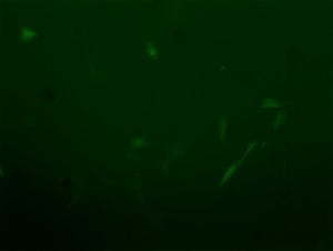
03/29/2018: Last week, I mentioned how the Osteopontin shRNA we used contained an antibiotic resistance. By treating the cells with that particular antibiotic, we could use the antibiotic to select cells that accepted the Osteopontin shRNA from the ones that didn’t. The Osteopontin shRNA also contained something called GFP (green fluorescent protein). Essentially, any cell that accepted the Osteopontin shRNA should not only have reduced Osteopontin and resistance to a specific antibiotic but will also glow green. I’m still in the process of confirming how much Osteopontin the ERMS and ARMS cells have after Osteopontin shRNA compared to the original cells. While we wait on those results though, here’s some further evidence that the cells accepted the Osteopontin shRNA: a picture of one of the ARMS cultures with beautiful green cells.
04/04/2018: We are collaborating with a colleague at Georgetown University to test the specificity of an Osteopontin inhibitor … one from a sea sponge! Western blots were also done using samples that contained high Osteopontin, no Osteopontin, or known amounts Osteopontin protein to validate/assess a multitude of different Osteopontin antibodies.
04/11/2018: After last week’s plethora of Western blots to validate/assess the many different Osteopontin antibodies we have, we’re at a bit of a careful-consideration point with the project. Each of these antibodies we tested seem to have useful and otherwise-complicating features (whether an antibody works for mouse, human or both; whether it recognizes Osteopontin correctly and/or proteins that are not Osteopontin), We need a best way to confirm how much Osteopontin the ERMS and ARMS cells have after Osteopontin shRNA compared to the original cells. Thus, we’re regrouping and figuring out the best strategy to confirm this before moving forward.
04/18/2018: After an Osteopontin team meeting, we’ve devised a plan forward for which antibody is best for which application – we will be evaluating Osteopontin cells this week. Meanwhile our collaborator is starting a screen for small molecule osteopontin inhibitors.
04/25/2018: I’m in the process of evaluating our Osteopontin knock down cells. Additionally, I started an experiment last week using a different approach to evaluate our Osteopontin antibodies… I have transduced cells with a tagged Osteopontin so that I can compare the Osteopontin antibody signal with the tag antibody signal, which we know is a well validated antibody, in order to make sure the signals we are seeing are actually Osteopontin and not something else.
05/03/2018: The additional approach to evaluate Osteopontin antibodies is still underway, as the tagged-Osteopontin transduced cells are still undergoing bacterial selection. We’re very close to being able to validate Osteopontin knockdown in our previously made cells and use these cells in mouse experiments!
05/10/2018: Selection of the cells expressing tagged-Osteopontin will finish by the end of the week and then I’ll do Western blots.
update 6/06/2018:
By popular demand, we are switching to less-frequent, more content-rich monthly posts. These will appear as new blog posts on our website. For exclusive! week to week updates of late-breaking results, email molly@cc-tdi.org to be subscribed. Thank you for supporting this rhabdomyosarcoma research!